Home » Without Label » Blank Diagram Of A Long Bone : Long Bone Anatomy | Human Anatomy Quiz - Quizizz - Structure of a long bone diagram posted by admin diagram system october 17 2018 0920 22 views a long bone diagram structure u anatomy and physiologyrhopentextbcca periosteum longjpg black and white long diagram unlabelled rhanatomyhumanchartscom long bone diagram blank.
Blank Diagram Of A Long Bone : Long Bone Anatomy | Human Anatomy Quiz - Quizizz - Structure of a long bone diagram posted by admin diagram system october 17 2018 0920 22 views a long bone diagram structure u anatomy and physiologyrhopentextbcca periosteum longjpg black and white long diagram unlabelled rhanatomyhumanchartscom long bone diagram blank.
Blank Diagram Of A Long Bone : Long Bone Anatomy | Human Anatomy Quiz - Quizizz - Structure of a long bone diagram posted by admin diagram system october 17 2018 0920 22 views a long bone diagram structure u anatomy and physiologyrhopentextbcca periosteum longjpg black and white long diagram unlabelled rhanatomyhumanchartscom long bone diagram blank.. Sectional diagram of a long bone. Long bone images stock photos vectors shutterstock from image.shutterstock.com. Diagram of hand bones pics photos bones of the hand labeled diagram print wiring. Cheek bone (zygoma) upper jaw (maxilla). There is a printable worksheet available for download here so you can take.
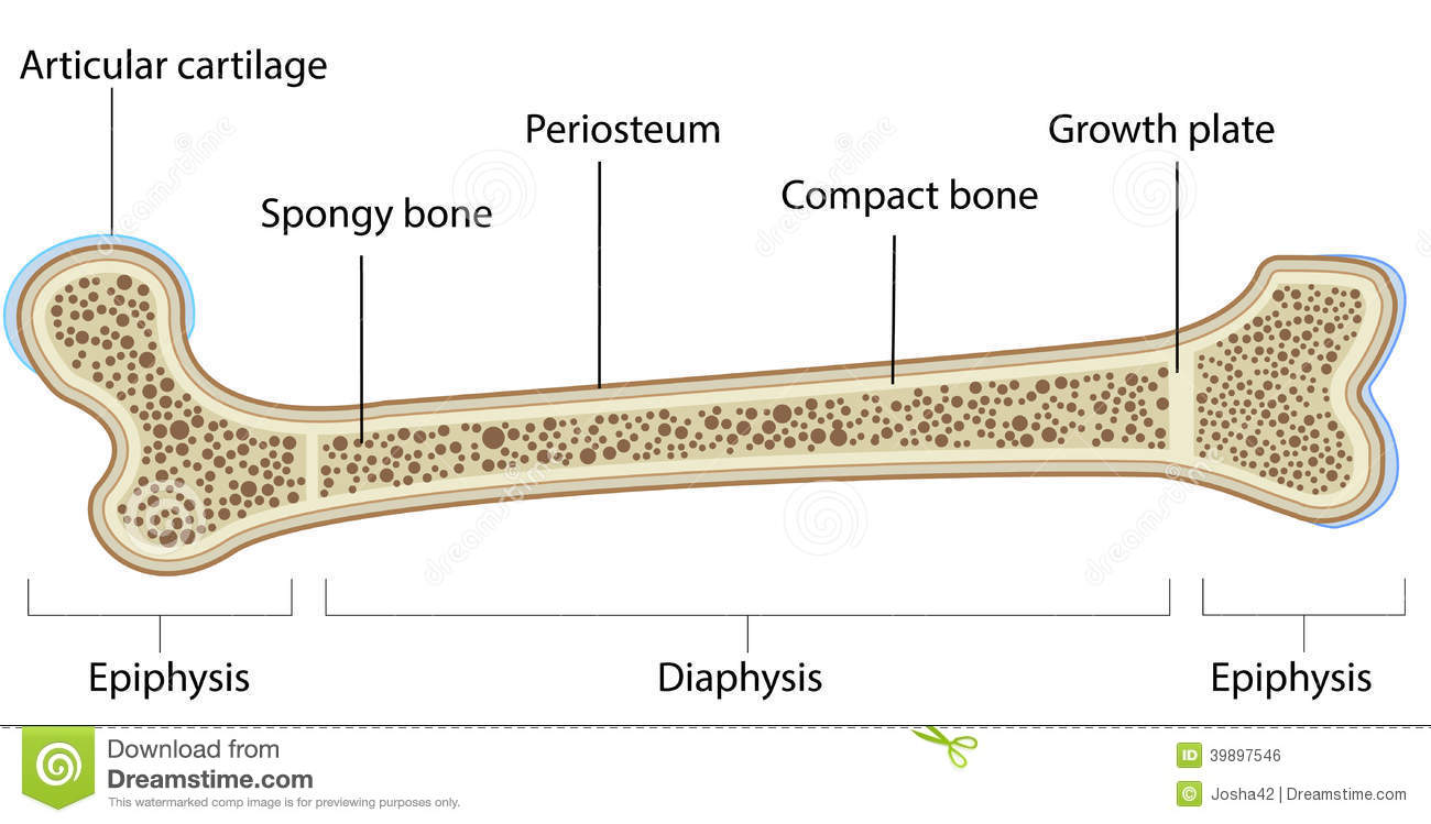
The mineral calcium phosphate hardens this framework, giving it strength. The long bones are those that are longer than they are wide. The hard cortical tissue can be invaded by cells that destroy the bone, called osteoclasts, only to have new bone laid down by secondary osteoblasts. Bone long diagram diaphysis tissue biology blood body cell compact humerus structure vector anatomical anatomy articular calcium cartilage detail education educational endosteum epiphysis forelimb health healthy human illustration joint long bone marrow medical. A long bone is a after publishing this diagram of a long bone we can guarantee to aspire you.
Long bone from www.askaboutireland.ie The hard cortical tissue can be invaded by cells that destroy the bone, called osteoclasts, only to have new bone laid down by secondary osteoblasts. Learn vocabulary terms and more with flashcards games and other study tools. Long bones, especially the femur and tibia, are subjected to most of the load during daily activities and they are crucial for skeletal mobility. Stranded diagram of a bone can be a kind of electrical wiring which happens to be greatly employed in properties. Supply the diaphysis and epiphysis. In long bones, chondrocytes form a template of the hyaline cartilage diaphysis. See that how can you get strong bone naturally. note the answers are at the bottom of this blog!
The outside of the bone consists of a layer of connective tissue.
Human anatomy for muscle reproductive and skeleton. Lower jaw (mandible) collar bone. There is a printable worksheet available for download here so you can take. End of bone furthest from the body's midline. Diagram of of a long bone. A labeled diagram of a long bone. See that how can you get strong bone naturally. The diaphysis is the tubular shaft that runs between the proxi. Diagram of a rat skeleton free wiring diagram for you. Structure of a long bone diagram posted by admin diagram system october 17 2018 0920 22 views a long bone diagram structure u anatomy and physiologyrhopentextbcca periosteum longjpg black and white long diagram unlabelled rhanatomyhumanchartscom long bone diagram blank. The hard cortical tissue can be invaded by cells that destroy the bone, called osteoclasts, only to have new bone laid down by secondary osteoblasts. The common name of each bone is listed first, with the scientific name given in parenthesis. The mineral calcium phosphate hardens this framework, giving it strength.
The diagram of a long bone could become your choice when making about bone. Download scientific diagram | 1 structure and components of long bone. Bones of the chest and upper back. End of bone furthest from the body's midline. See that how can you get strong bone naturally.
Skeleton Worksheet - WikiEducator from wikieducator.org Blank bone labeling blank bone diagram skull bones blank long bone drawing long bone model femur skeleton long bone worksheet long bone with labels long bone parts blank bone diagrams blank ulna blank skull blank heart blank humerus blank human torso bones. Download scientific diagram | 1 structure and components of long bone. Anatomy of a long bone anna s anatomy websit. The diaphysis is the tubular shaft that runs between the proxi. In various parts over the review. Learn vocabulary terms and more with flashcards games and other study tools. Bone long diagram diaphysis tissue biology blood body cell compact humerus structure vector anatomical anatomy articular calcium cartilage detail education educational endosteum epiphysis forelimb health healthy human illustration joint long bone marrow medical. Diagram of hand bones pics photos bones of the hand labeled diagram print wiring.
When a human finishes growing these parts fuse together.
When a human finishes growing these parts fuse together. Membranous lining of the hollow cavity of the bone. During the course of development, the bone tissue is recycled, gradually altering its shape. The mineral calcium phosphate hardens this framework, giving it strength. The long bones are those that are longer than they are wide. Not only blank bone diagram, you could also find another pics such as blank hand diagram, human skeleton diagram blank, blank foot diagram, blank anatomy diagram, blank. Bone is found in the shafts of long bone and consists of various cylindrical units named as haversian system 47. Long bones — a subtype of bones — are longer than they are wide. In long bones, chondrocytes form a template of the hyaline cartilage diaphysis. Find out where this is usually located and, if it is present, label it on your bone. Long blank long bone diagram bone structure diagram and metaphysisjpg from the above resolutions which is part of the human anatomydownload this image for free in hd resolution the choice download button diagram of a long bone. Learn vocabulary terms and more with flashcards games and other study tools. They are one of five types of bones:
Structure of a long bone diagram posted by admin diagram system october 17 2018 0920 22 views a long bone diagram structure u anatomy and physiologyrhopentextbcca periosteum longjpg black and white long diagram unlabelled rhanatomyhumanchartscom long bone diagram blank. Long bones may suffer from different types of fractures. Long, short, flat, irregular and sesamoid. Sectional diagram of a long bone. While their parts are similar in general, their structure has.
Periosteum clipart 20 free Cliparts | Download images on ... from clipground.com The diagram of a long bone could become your choice when making about bone. Stranded diagram of a bone can be a kind of electrical wiring which happens to be greatly employed in properties. Bones of the chest and upper back. General features of a long bone file human arm bones diagram svg anatomy of long bone Human anatomy for muscle reproductive and skeleton. Bone is found in the shafts of long bone and consists of various cylindrical units named as haversian system 47. Diagram of of a long bone. The hard cortical tissue can be invaded by cells that destroy the bone, called osteoclasts, only to have new bone laid down by secondary osteoblasts.
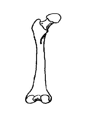
The long bone diagram blank could be your desire when thinking of about bone.
While their parts are similar in general, their structure has. The hard cortical tissue can be invaded by cells that destroy the bone, called osteoclasts, only to have new bone laid down by secondary osteoblasts. Sectional diagram of a long bone. Long bone images stock photos vectors shutterstock from image.shutterstock.com. The end of the long bone is the epiphysis and the shaft is the diaphysis. Your drawing should be in pencil. The long bones are those that are longer than they are wide. Each system bone tissue engineering is currently a hot research field and aims to realize bone reconstruction and regeneration, focusing on scaffolds, cells, growth. File axial skeleton diagram blank svg wikimedia commons. Fibula, outer of two bones of the lower leg or hind limb. Find out where this is usually located and, if it is present, label it on your bone. They are one of five types of bones: Cheek bone (zygoma) upper jaw (maxilla).